What is MRI?
MRI is an acronym for magnetic resonance imaging, and it is a technology which uses a combination of strong magnets and radio waves to produce internal images of the body. The MRI machine is a large, hollow magnet. During the scan, the patient lies on a bed which moves horizontally into the opening of the magnet. The body region being studied is generally placed in the middle of the machine. Using the interactions of coils, magnets, and radio waves, MRI generates extremely detailed and accurate 3D images of the body.
MRI is highly useful in a number of diagnoses and treatments. It is often used to scan sporting injuries, brain and spinal cord, heart, blood vessels, and internal organs of the abdomen, pelvis, and chest. Your physician, in conjunction with your radiologist, can advise on the best and most effective scan for your particular health concern.
Magnetic resonance imaging (MRI) is a medical imaging technique that uses a magnetic field and computer-generated radio waves to create detailed images of the organs and tissues in your body.
Most MRI machines are large, tube-shaped magnets. When you lie inside an MRI machine, the magnetic field temporarily realigns water molecules in your body. Radio waves cause these aligned atoms to produce faint signals, which are used to create cross-sectional MRI images — like slices in a loaf of bread.
The MRI machine can also produce 3D images that can be viewed from different angles.
MRI is a safe and non-invasive procedure, and it is one of the most common radiological tests we perform at Vision XRAY. Unlike traditional x-ray or CT scan (other common procedures) the MRI does not use any ionising radiation.

Why it’s done
MRI is a noninvasive way for your doctor to examine your organs, tissues and skeletal system. It produces high-resolution images of the inside of the body that help diagnose a variety of problems.
What to Expect When Getting an MRI
Although getting a magnetic resonance imaging scan is not a fun thing to do, sometimes it’s a medical necessity. Most individuals want to know what they can expect before they get their first MRI, as it can be unnerving when you don’t know what’s going to happen.
While it’s normal and common to have anxiety before an MRI, you won’t be as anxious when you know what to expect and how to prepare.
What is an MRI Appointment Like?

An MRI does not typically require excessive preparation. Usually, you are permitted to eat and drink as usual during the days prior to your appointment. You may be instructed to avoid food or beverage for a short time period (such as 30 minutes) leading up to your scan. Upon arrival, you will need to remove any items which might interfere with the magnetic imaging, such as jewellery and other metallic items. It can often be easier to simply leave these items at home. You will change into a gown and it will be time for the scan.
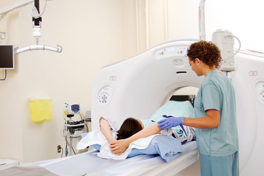
Usually, the scan lasts for half an hour to an hour, depending upon the area of the body under study. Whilst in the machine, you will need to lie still during the scan and may occasionally be asked to hold your breath for several seconds. You will be able to communicate with the radiographer through headphones, which can help you feel more comfortable and at ease. The MRI machine can sometimes create loud knocking sounds.
Sometimes, a contrast dye is required for an MRI to better illuminate internal organs and bring details into sharp focus on imaging. This is usually administered via an injection. Prior to your appointment, we will discuss if this will be necessary for your test and the decision is always up to you.
Before the scan
On the day of your MRI scan, you should be able to eat, drink and take any medication as usual, unless you’re advised otherwise.
In some cases, you may be asked not to eat or drink anything for up to 4 hours before the scan, and sometimes you may be asked to drink a fairly large amount of water beforehand. This depends on the area being scanned.
When you arrive at the hospital, you’ll usually be asked to fill in a questionnaire about your health and medical history. This helps the medical staff ensure you have the scan safely.
Once you have completed the questionnaire, you’ll usually be asked to give your signed consent for the scan to go ahead.
As the MRI scanner produces strong magnetic fields, it’s important to remove any metal objects from your body.
These include:
- watches
- jewellery, such as rings and necklaces
- piercings, such as ear, nipple and nose rings
- dentures (false teeth)
- hearing aids
- wigs (some wigs contain traces of metal)
It’s best not to bring any valuables with you to your scan, but if you do they can usually be stored in a secure locker.
Depending on which part of your body is being scanned, you may need to wear a hospital gown during the procedure.
If you don’t need to wear a gown, you should wear clothes without metal zips, fasteners, buttons, underwire (bras), belts or buckles.
Contrast agent
Some MRI scans involve having an injection of contrast agent (dye). This makes certain tissues and blood vessels show up more clearly and in greater detail.
Sometimes the contrast agent can cause side effects, such as:
- feeling or being sick
- a skin rash
- a headache
- dizziness
These side effects are usually mild and don’t last very long.
It’s also possible for contrast agent to cause tissue and organ damage in people with severe kidney disease.
If you have a history of kidney disease, you may be given a blood test to determine how well your kidneys are functioning and whether it’s safe to proceed with the scan.
You should let the staff know if you have a history of allergic reactions or any blood clotting problems before having the injection.
Anaesthesia and sedatives
An MRI scan is a painless procedure, so anaesthesia (painkilling medication) isn’t usually needed.
If you’re claustrophobic, you can ask for a mild sedative to help you relax. You should ask your GP or consultant well in advance of having the scan.
If you decide to have a sedative during the scan, you’ll need to arrange for a friend or family member to drive you home afterwards, as you won’t be able to drive for 24 hours.
Babies and young children may be given a general anaesthetic before having an MRI scan.
This is because it’s very important to stay still during the scan, which babies and young children are often unable to do when they’re awake.
During the scan
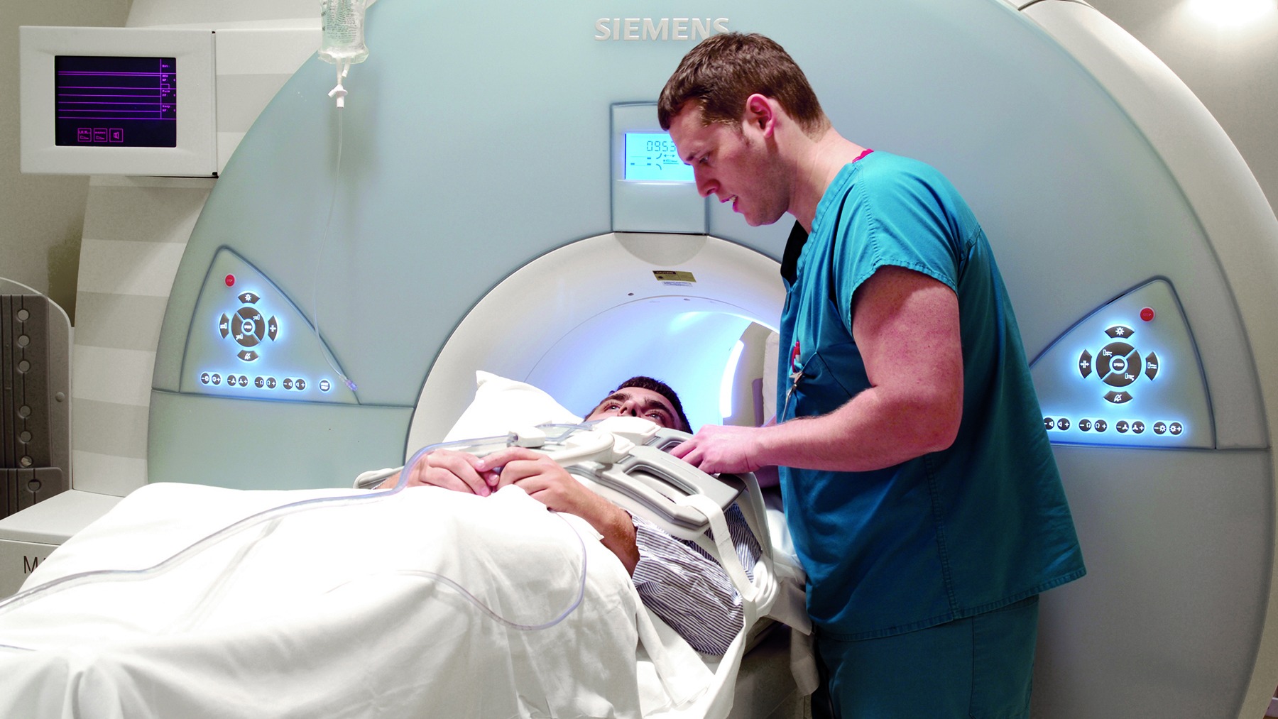
An MRI scanner is a short cylinder that’s open at both ends. You’ll lie on a motorised bed that’s moved inside the scanner.
You’ll enter the scanner either head first or feet first, depending on the part of your body being scanned.
In some cases, a frame may be placed over the body part being scanned, such as the head or chest.
This frame contains receivers that pick up the signals sent out by your body during the scan and it can help to create a better-quality image.
A computer is used to operate the MRI scanner, which is located in a different room to keep it away from the magnetic field generated by the scanner.
The radiographer operates the computer, so they’ll also be in a separate room to you.
But you’ll be able to talk to them, usually through an intercom, and they’ll be able to see you at all times on a television monitor and through the viewing window throughout the scan.
A friend or family member may be allowed to stay with you while you’re having your scan. Children can usually have a parent with them.
Anyone who stays with you will be asked if they have a pacemaker or any other metal objects in their body.
They’ll also have to follow the same guidelines regarding clothing and the removal of metallic objects.
To avoid the images being blurred, it’s very important to keep your whole body still throughout the scan until the radiographer tells you to relax.
A single scan may take from a few seconds to 3 or 4 minutes. You may be asked to hold your breath during short scans.
Depending on the size of the area being scanned and how many images are taken, the whole procedure will take 15 to 90 minutes.
The MRI scanner will make loud tapping noises at certain times during the procedure. This is the electric current in the scanner coils being turned on and off. You’ll be given earplugs or headphones to wear.
You’re usually able to listen to music through headphones during the scan if you want to, and in some cases you can bring your own CD.
You’ll be moved out of the scanner when your scan is over.
After the scan
An MRI scan is usually carried out as an outpatient procedure. This means you won’t need to stay in hospital overnight.
After the scan, you can resume normal activities immediately. But if you have had a sedative, a friend or relative will need to take you home and stay with you for the first 24 hours.
It’s not safe to drive, operate heavy machinery or drink alcohol for 24 hours after having a sedative.
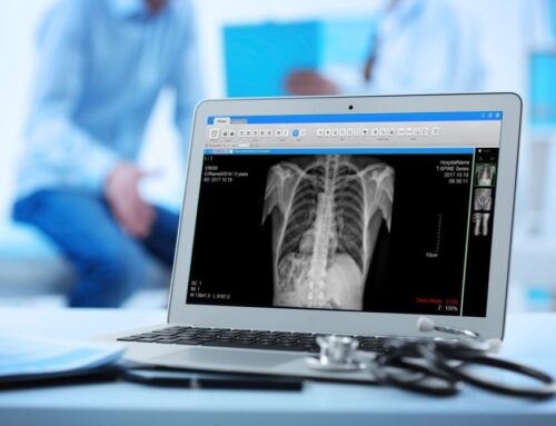
Your MRI scan needs to be studied by a radiologist (a doctor trained in interpreting scans and X-rays) and possibly discussed with other specialists.
This means it’s unlikely you’ll get the results of your scan immediately.
The radiologist will send a report to the doctor who arranged the scan, who will discuss the results with you.
It usually takes a week or two for the results of an MRI scan to come through, unless they’re needed urgently.
If you feel pain or any unusual symptom following the exam, contact your referring physician. You can be as active as you like after the MRI unless you were given a sedative. Check with your doctor about this. The pictures taken during the test will be reviewed by a radiologist. Your results will then be given to your doctor, who will discuss them with you.

MRI of the brain and spinal cord
MRI is the most frequently used imaging test of the brain and spinal cord. It’s often performed to help diagnose:
- Aneurysms of cerebral vessels
- Disorders of the eye and inner ear
- Multiple sclerosis
- Spinal cord disorders
- Stroke
- Tumors
- Brain injury from trauma
A special type of MRI is the functional MRI of the brain (fMRI). It produces images of blood flow to certain areas of the brain. It can be used to examine the brain’s anatomy and determine which parts of the brain are handling critical functions.
This helps identify important language and movement control areas in the brains of people being considered for brain surgery. Functional MRI can also be used to assess damage from a head injury or from disorders such as Alzheimer’s disease.
MRI of the heart and blood vessels
MRI that focuses on the heart or blood vessels can assess:
- Size and function of the heart’s chambers
- Thickness and movement of the walls of the heart
- Extent of damage caused by heart attacks or heart disease
- Structural problems in the aorta, such as aneurysms or dissections
- Inflammation or blockages in the blood vessels
MRI of other internal organs
MRI can check for tumors or other abnormalities of many organs in the body, including the following:
- Liver and bile ducts
- Kidneys
- Spleen
- Pancreas
- Uterus
- Ovaries
- Prostate
MRI of bones and joints
MRI can help evaluate:
- Joint abnormalities caused by traumatic or repetitive injuries, such as torn cartilage or ligaments
- Disk abnormalities in the spine
- Bone infections
- Tumors of the bones and soft tissues
MRI of the breasts
MRI can be used with mammography to detect breast cancer, particularly in women who have dense breast tissue or who might be at high risk of the disease.
What is an MRI scan used to diagnose?
MRI has proven valuable in diagnosing a broad range of conditions, including cancer, heart and vascular disease, and muscular and bone abnormalities. MRI can detect abnormalities that might be obscured by bone with other imaging methods.
Is MRI scan painful?
An MRI scan is a painless procedure, so anaesthesia (painkilling medication) isn't usually needed. If you're claustrophobic, you can ask for a mild sedative to help you relax. You should ask your GP or consultant well in advance of having the scan.

