One of the topmost results of the innumerous advancements in radiological technology is the growth in dental imaging. We offer a variety of options in dental imaging, aiding dental interpreters in their work and effective treatment of cases. With our range of outfit, we can produce specialised, high quality images for use by dental and medical providers, examining not only the teeth and jaw, but also imaging facial bones, dental jitters, and more.
Digital Radiography
For further simple images of the mouth and teeth, we use first class 2- D digital radiography. These low cure machines produce excellent images for use by your dental provider. We perform side cephalometry, OPG, TMJ, and facial bone imaging using this technology.
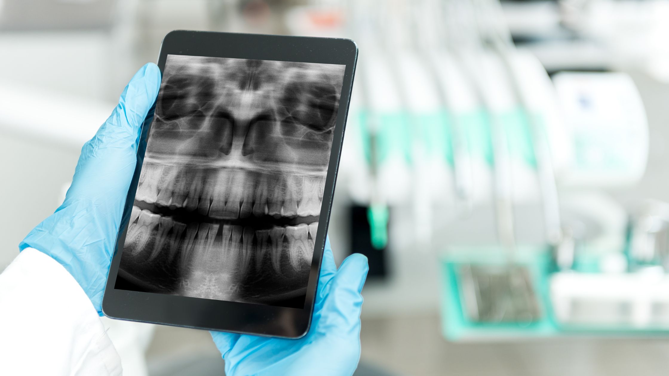
OPG (orthopantomogram) provides a panoramicx-ray of the case’s lower face, imaging the teeth of both the upper and lower jaw. This checkup can be used to display the position and growth of all teeth, including those that are still to face, and may also be used to examine the structure of the jawbone and the temporomandibular joint (TMJ). OPG’s are constantly used when checking for the presence of wisdom teeth in a case.
Side cephalometry is anx-ray which produces a side view of the face, showing bone and facial structure. Such a checkup is useful for orthodontists in developing their treatment plans and tracking progress.
Cone Ray CT
Our cone ray CT( computerized tomography) is an ultra-low cure dental CT checkup. This procedure uses advanced technology to produce 3- D images of the craniofacial region. An cone shapedx-ray ray is moved around the head of the case to induce clear images of dental structures, jitters, soft apkins, and bone. Because these images are more detailed, cone ray CT can help in more specific and specialised treatment and opinion.
Cone ray CT may be helpful for orthodontic treatment as well as surgical planning for issues similar as teeth impaction. TMJ disorder opinion can also be supported through this technology, and issues with the jaw, sinuses, and nasal depression can also be estimated. When cases are passing pain in the craniofacial region, the cone ray CT can be necessary in abetting radiologists to determine the source of the problem. One of the most significant uses for cone ray CT is its part in planning tooth implantation. Cone ray CT shows the height and range of bone available for implantation as well as bone quality. This assists in the planning the procedure as well as determining whether or not the procedure can be safely performed.