X-ray. CT scan. Ultrasound. MRI. To a patient, sometimes these terms can sound overwhelming and meaningless. We strive to ensure all patients have a better understanding of the various procedures and treatments we offer at our practices. The equipment and scanning options we use make up the radiologist’s toolbox; each is a valuable resource, useful and effective for various conditions, injuries, and circumstances. Today, we present to you a basic guide to our most common procedures.
XRAY
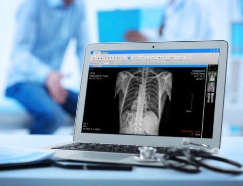
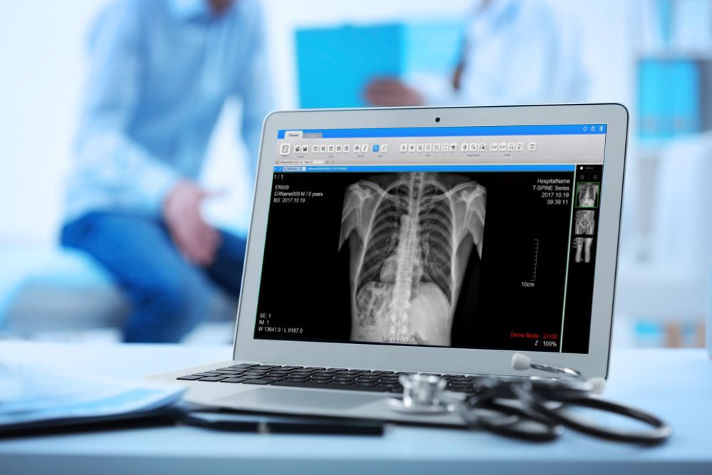
The x-ray is a frequently used procedure in our offices. The x-ray is painless and fast, and produces images of internal structures inside your body. X-rays use ionising radiation, which pass through your body to create the images. Structures inside your body absorb the rays differently, and show up as gradients of black and white in the image. Bones appear white because the calcium inside them is highly absorbent to the waves. This type of test is commonly used to observe broken bones, to examine the lungs, to imagine the teeth, or in mammogram, a specialised x-ray which images the breast tissue for abnormalities.
CT
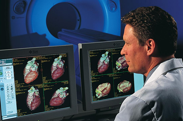
The computerised tomography scan, or CT, is a type of x-ray which rotates around the patient’s body to build cross-section, 3D images of that area. Also a pain free procedure, the CT is fairly quick and may involve the ingestion of a liquid or an injection of a contrast dye, to help produce better images. The CT is an excellent tool for examining internal injuries or scanning for abnormalities. Because the CT generates an image of finer detail, it is often preferred over the basic x-ray. It is also able to be localised onto a specific area, to better focus the scan and to minimise radiation exposure.
MRI
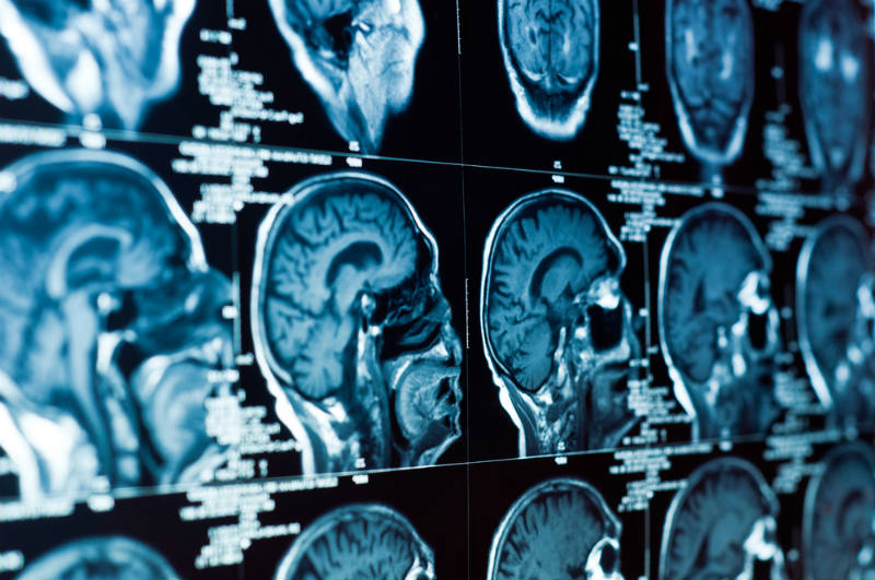
MRI stands for magnetic resonance imaging, a scan which utilises radio waves and a magnet to generate remarkably intricate images of a patient’s body. The MRI machinery uses wire coils to transmit radio waves and the resulting signals receive build a series of cross-section images, creating a multi-dimensional understanding of the bodily tissues. MRI’s are often selected to scan tissues or areas in greater detail, such as ligaments, tendons, or the spinal cord.
An MRI lasts a bit longer than a CT scan or x-ray, can be quite noisy and may require the use of an orally-ingested or IV contrast dye to assist with imaging.
ULTRASOUND
Ultrasound is a safe type of scan which utilises penetrating sound waves to present images of the body. Women who have had babies will know the ultrasound as a common tool seen over the course of pregnancy. Ultrasound technology allows the radiologist and physician to monitor and observe the health of the growing foetus and provides excellent detail. Ultrasound is also used for other purposes and with other areas of the body, including breast ultrasound, pelvic ultrasound, vascular ultrasound and musculoskeletal ultrasound. Effective in determining the mass and type of abnormalities within the body, ultrasound is a useful part of diagnosis, often as a supplementary tool alongside other imaging tests.
What does an ultrasound check for?
The test can provide information about a baby's growth, development, and overall health. Diagnostic ultrasound is used to view and provide information about other internal parts of the body. These include the heart, blood vessels, liver, bladder, kidneys, and female reproductive organs.
What does a CT scan check for?
CT scans can detect bone and joint problems, like complex bone fractures and tumors. If you have a condition like cancer, heart disease, emphysema, or liver masses, CT scans can spot it or help doctors see any changes. They show internal injuries and bleeding, such as those caused by a car accident.