Every day, advances in radiology show just how far science and technology have brought us. Within the medical field, progress in radiology has always been at the forefront, and it continues its amazing growth today. While we typically focus on present-day radiological technologies, it is fascinating to peer into the past and see just how far we’ve come. Join us as we explore a brief history of a modern marvel: radiology.
The Beginnings
In 1895, Wilhelm Conrad Rontgen discovered the X-ray. Rontgen was experimenting with the flow of an electric current in a glass tube (cathode-ray tube) when he noticed that a nearby piece of barium platinocyanide emitted light during his experiment. He proposed that when the cathode rays (electrons) encountered the wall inside the tube a type of radiation formed which moved across the room and caused the illumination. He called this X-radiation, or Rontgen radiation, and found that the radiation worked transparently with materials such as paper and aluminum. Rontgen is responsible for taking the very first X-ray photographs, producing images of metal objects as well as scanning the bones in his wife’s hand
Developments
The X-ray apparatus the Rontgen created was widely available, so many scientists and physicians were able to work with and study the technology. In the 1910’s, many physicians purchased their own x-ray machines. As time went on, radiology transformed into a specialist profession, separate from a physician.

The earliest x-ray images were made onto glass photographic plates, but in 1918 film was introduced by George Eastman. This allowed for more efficient (and likely less costly) x-ray photographs. Study of radiology continued throughout the 20th century, and by 1920, the Society of Radiographers was formed.
Advances in the technology helped improve the speed and efficacy of X-rays, and their use was broadened. In the 1940’s and 50’s, more safety regulations and precautions were understood and put in place. X-rays became increasingly useful in treatment and diagnosis, and could assist with more serious issues.
The 1950’s and 60’s saw the development and spread of ultrasound technology. With continued growth, radiology was suddenly booming, and there was a shortage of radiologists in the late 1960’s and early 70’s. Scanning procedures such as computed tomography (CT) and mammography and were becoming increasingly common. During this period, radiological societies pushed for more standardisation of radiological training and education, to ensure professionals were always qualified and expertly trained. and for higher Fearing that the shortage would lead to “diploma mills” that churned out
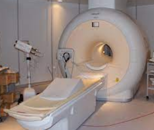
CT scanning was developed in the early 70’s by Englishman Godfrey Hounsfield and the first CT scan performed in 1972; he won the Nobel Prize for his work. MRI (magnetic resonance imaging) was also under exploration, and the first image of a human was completed in 1977 in Scotland. Work was progressing on magnetic resonance imaging in the 1970s and the first human image was obtained in Aberdeen in 1977. Finally, radiology offered a myriad of options for patients, leading to more accurate diagnosis and better treatment.
Leading Up to Today
The 1970’s and 80’s saw an important growth in technology: the computer. These achievements were revolutionary, as computerised x-rays could provide dramatically better imagery and information. Beyond the common x-ray modern radiology works with others sources of energy: ultrasound waves, isotopes, and an an electromagnetic field, (MRI). Computerisation works tremendously alongside these technologies to improve processes. The 80’s and 90’s also gave birth to teleradiology, allowing radiologists, physicians, and health centres to communicate and share patient images easily and swiftly via computers, scanners, and other equipment, This has allowed for amazing collaboration between radiologists and physicians, and resulted in overall better patient care.
What is the history of radiography?
X-rays were discovered in 1895 by Wilhelm Conrad Roentgen (1845-1923) who was a Professor at Wuerzburg University in Germany. Working with a cathode-ray tube in his laboratory, Roentgen observed a fluorescent glow of crystals on a table near his tube.
Why is it called radiology?
Radiographs (originally called roentgenographs, named after the discoverer of X-rays, Wilhelm Conrad Röntgen) are produced by transmitting X-rays through a patient.

