Back pain is a common reason people miss work or visit their doctor. According to the National Institutes of Health (NIH), about 80% of adults suffer from lower back pain at one point or another. Back problems can make it difficult to enjoy life and carry out your regular daily activities. The good news is, most back pain is treatable without surgery, and medical professionals are here to help.
The first step to treating back pain is to find the cause. Your doctor may order an imaging test to determine the reason for your back discomfort. Tests such as x-rays, CT scans and MRIs allow a doctor to look into your back without performing surgery. Imaging tests are quick, painless procedures that provide valuable insight into your condition. We’ll look at the types of imaging tests commonly used to diagnose back pain, but first, it helps to understand what causes back discomfort. That way, you’ll know why your doctor ordered a specific test.
Common Causes of Back Pain
There are two types of back pain: acute and chronic. Acute back pain occurs suddenly and may last up to six weeks. Chronic back pain is less common and lasts longer than three months.
Many times, back pain begins without a known cause. An imaging test allows your doctor to find the cause of your pain, so that they can treat the issue effectively. Some possible common causes of back pain include:
- Muscle strains or ligament sprains: Strains and sprains are the most common causes of acute back pain. Both of these occur from picking up heavy objects, over-stretching or lifting something improperly. A strain or sprain can lead to painful spasms in the back.
- Disc degeneration: If you were to look between the vertebrae, or bones in your spine, you’d find rubbery discs. These intervertebral discs act as cushions to absorb shocks and help you bend your back. Over time, these discs can deteriorate and become more prone to rupturing or tearing. A bulging or ruptured disc might press on a nerve and cause back pain.
- Spinal stenosis: Spinal stenosis is the narrowing of the space around your spinal cord. This condition often affects the lower back and neck and may result in pain or numbness. It’s usually caused by spinal changes related to osteoarthritis.
- Skeletal irregularities: Skeletal irregularities such as scoliosis, which is the curvature of the spine, may cause back pain. However, scoliosis usually does not cause pain until middle age.
- Osteoporosis: Osteoporosis is a disease that causes bones to weaken. Individuals with osteoporosis are more vulnerable to experiencing fractures in their vertebrae.
- Traumatic injury: Playing sports, falling or being in a car accident may lead to injured muscles or ligaments in the back or a ruptured disc. Any of this could result in back pain.
Back pain is rarely a sign of a severe underlying condition, but it can be. A complete physical exam and imaging test for back pain will help your doctor pinpoint the cause so you can get the attention you need.
Types of Tests Used for Diagnosing Back Pain
Imaging tests create pictures of the inside of your body. They provide clues to help your doctor find out the source of your back irritation. Your doctor will select a test depending on your symptoms and the location of your back pain. Common types of scans used to diagnose back issues include the following.
CT Scan for Back Pain
![]()
80% of all Australians will suffer from back pain at some time in their lives and 1 in 10 people are limited in their activity caused by back pain.
Back pain is the most significant work-related problem in Australia.
It is a much more common long-term condition than asthma, arthritis or high blood pressure.
Back pain is not one single disease and has many causes. Some are related to the structure of the spine like joints, discs and ligaments and are brought on by lifting, carrying, pushing or pulling loads that are too heavy or going about these tasks in an unsafe manner. Back issues and pain can also be related to standing, sitting or bending down for long periods, having a trip or a fall, being overweight or having poor posture.
There may be other, more serious but uncommon underlying causes of your back pain such as fracture, osteoporosis, a slipped disc or arthritis in a joint of the back.
Most of the time back pain gets better by itself or is relieved by painkillers. If the back pain lasts for more than 3 months it is then considered as chronic and treatment should be commenced immediately.
Pinpointing the problems that cause back pain is the most difficult task for doctors. MRI or CT scan in combination with your doctors understanding of your symptoms is most important to understand the abnormality.
Depending on severity, treatment can range all the way from a physical therapy routine you do do at home, to surgery to solve the problem.
We see patients referred by other doctors for severe back pain management. This is done using a CT scan as a guide to the right spot. Then drugs are placed into deep parts of the back to relieve pain coming from nerves and from joints. These treatments are called peri-radicular blocks or facet joint blocks. They do not cure the problem and are used for medium to long-term pain relief in chronic back pain so that you can live a normal work and home life.
Any time CT scans are used, it is important to make sure that the lowest radiation dose possible is given.
Your doctor might order a CT scan to diagnose a back injury related to soft tissues. A CT scan shows images of muscles, organs and ligaments more clearly than a conventional x-ray. CT scans are also often used in response to an emergency such as an accident or other trauma.
A CT scan machine produces multiple x-rays at different angles to create a detailed 3-D image of your bones and soft tissues. It can be an extremely useful diagnostic tool and helps prevent the need for exploratory surgery. A CT scan may be used to diagnose the following:
- Disc degeneration
- Ruptured discs
- Spinal stenosis
- Spinal cord infections or tumors
Today’s CT scanners are faster than ever and complete a scan within minutes. At an Envision Imaging location, you can expect state-of-the-art CT scan equipment and technologists who care about your comfort and safety.
2. X-Ray for Back Pain
An x-ray machine uses electromagnetic radiation to capture images of the inside of your body. Dense materials such as bones and metal appear white in an x-ray image, while muscles, fat and fluids appear dark. Your doctor may order an x-ray for back pain to check for the following:
- Fractured vertebrae
- Spinal changes due to osteoarthritis
- Spine alignment issues
X-rays are usually the first type of test a doctor will order to rule out bone injuries. Getting a back x-ray is a quick, painless and noninvasive procedure. Your doctor will need to perform a different imaging test if they suspect issues with the muscles, nerves or discs in your back.
If you get an x-ray for back pain at an Envision Imaging center, you can expect a specially trained technologist to perform the procedure as efficiently as possible and make sure you are comfortable throughout.
3. MRI for Back Pain
MRIs use magnetic fields and radio waves to create highly detailed images of your spine, muscles, bones, organs and other tissues. A doctor might order an MRI for a back pain diagnosis if they suspect any of the following:
- Infection
- Tumor
- Disc rupture
Like many other forms of diagnostic imaging, MRIs are a painless, noninvasive way for a doctor to see clearly into your back and find the source of your pain. Some patients worry they will feel claustrophobic or anxious during an MRI. At Envision Imaging, we use the latest MRI technology, which is not as restricting as outdated equipment. In addition, our technologists are trained to help you relax and feel as comfortable as possible during the procedure.
MRI vs. X-Ray for Back Pain
An MRI and x-ray are both painless procedures and valuable diagnostic tools, and each one has benefits. For example, an MRI is a much better device for diagnosing soft tissue problems, and it does not expose patients to ionizing radiation. An x-ray mainly helps doctors identify bone issues and is a faster procedure than an MRI.
Your doctor will discuss the best imaging option for you as well as the risks and benefits.
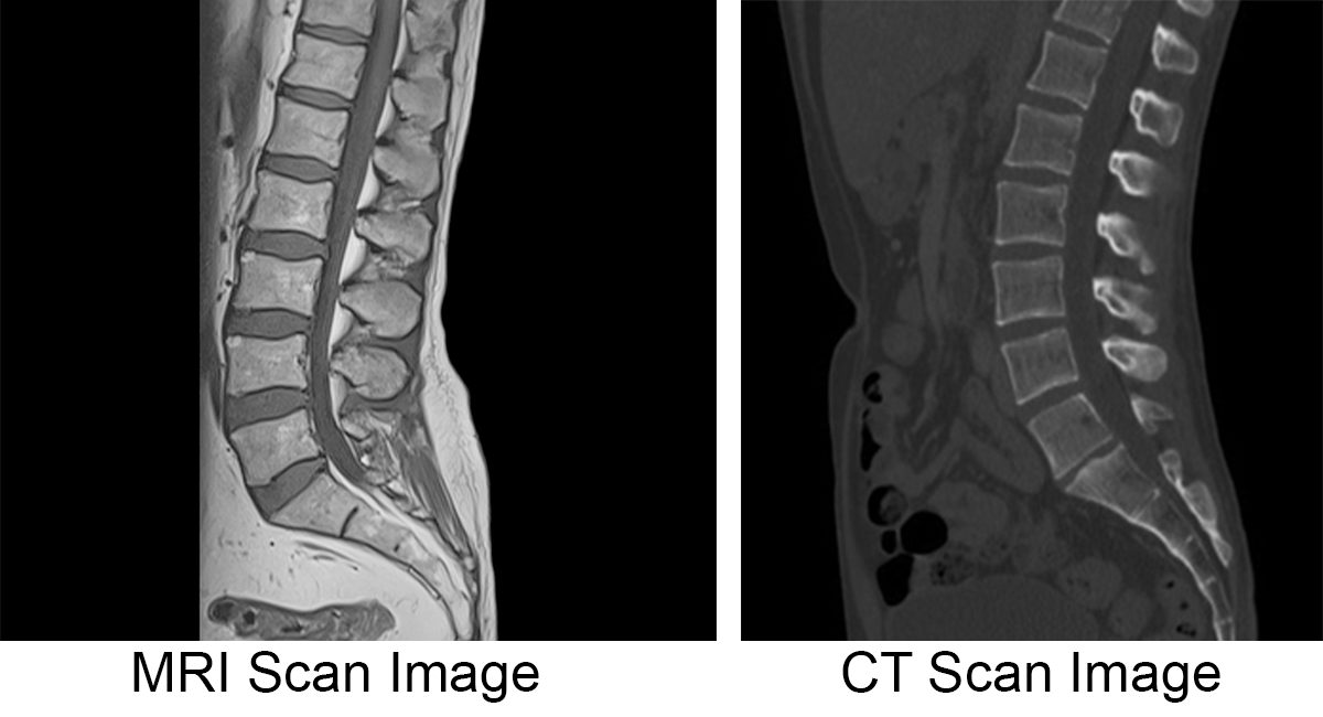
CT Scan vs. MRI for Back Pain

Like x-rays, CT scans are usually quicker than MRIs. CT scans are the preferred tool for diagnosing severe injuries that need immediate attention, and they are also helpful in locating tumors. Typically, CT scans are better at scanning bone images than MRIs. However, they expose patients to a small dose of radiation, whereas MRIs do not. MRIs are ideal for diagnosing soft tissue and spinal ligament issues.
The tests will not help you feel better faster.
Most people with lower-back pain feel better in about a month, whether or not they have an imaging test.
People who get an imaging test for their back pain do not get better faster. And sometimes they feel worse than people who took over-the-counter pain medicine and followed simple steps, like walking, to help their pain.
Imaging tests can also lead to surgery and other treatments that you do not need. In one study, people who had an MRI were much more likely to have surgery than people who did not have an MRI. But the surgery did not help them get better any faster.
Imaging test have risks.
X-rays and CT scans use radiation. Radiation has harmful effects that can add up. It is best to avoid radiation when you can.
Imaging tests are expensive.
Imaging tests can costs hundreds, or even thousands, of dollars depending on the test and where you have it. Why waste money on tests when they don’t help your pain? And if the tests lead to surgery, the costs can be much higher.
When are imaging tests a good idea?
In some cases you may need an imaging test right away. Talk to your doctor if you have back pain with any of the following symptoms:
- Weight loss that you cannot explain
- Fever over 102° F
- Loss of control of your bowel or bladder
- Loss of feeling or strength in your legs
- Problems with your reflexes
- A history of cancer
These symptoms can be signs of nerve damage or a serious problem such as cancer or an infection in the spine.
If you do not have any of these symptoms, we recommend waiting a few weeks.
How Do I Know What Kind of Back Problem I Have?
Unless you are totally immobilized from a back injury, your doctor probably will examine your range of motion and nerve function and touch your body to locate the area of discomfort.
Blood and urine tests may be done to determine if the pain is caused by an infection or other systemic problem.
X-rays are useful in pinpointing broken bones or other skeletal defects. They can sometimes help locate problems in connective tissue. To analyze soft-tissue or disc damage, computed tomography (CT) or magnetic resonance imaging (MRI) scans may be needed. X-rays and imaging studies are generally used to confirm your symptoms and exam results to identify the source of pain. Scans are also utilized in cases of direct trauma to the back, back pain with fever, or weakness or numbness in the limbs. To determine possible nerve or muscle damage, an electromyogram (EMG) may be ordered.
Is a CT scan good for back pain?
Your doctor might order a CT scan to diagnose a back injury related to soft tissues. A CT scan shows images of muscles, organs and ligaments more clearly than a conventional x-ray. CT scans are also often used in response to an emergency such as an accident or other trauma.
Is CT or MRI better for back?
Magnetic resonance imaging produces clearer images compared to a CT scan. In instances when doctors need a view of soft tissues, an MRI is a better option than x-rays or CTs. MRIs can create better pictures of organs and soft tissues, such as torn ligaments and herniated discs, compared to CT images.
Why would a doctor order a CT scan of the back?
A CT scan of the spine may be performed to assess the spine for a herniated disk, tumors and other lesions, the extent of injuries, structural anomalies such as spina bifida (a type of congenital defect of the spine), blood vessel malformations, or other conditions, particularly when another type of examination, such ...
Which scan is best for back pain?
You will likely need an MRI right away if you have warning signs of a more serious cause of back pain: Fever. History of cancer. Other signs or symptoms of cancer. Recent serious fall or injury. Back pain that is very severe, and not even pain pills from your provider help. One leg feels numb or weak and it is getting worse.
Can a CT scan show inflammation of the spine?
A scan of the spine may also be done after injecting contrast material into the spinal canal (usually well below the bottom of the spinal cord) during a lumbar puncture test, also known as a myelogram. This will help to locate areas of inflammation or nerve compression or detect tumors.
What are the red flags of back pain?
“Red flags” include pain that lasts more than 6 weeks; pain in persons younger than 18 years or older than 50 years; pain that radiates below the knee; a history of major trauma; constitutional symptoms; atypical pain (eg, that which occurs at night or that is unrelenting); the presence of a severe or rapidly ...
What is a drawback to using a CT scan?
Concerns about CT scans include the risks from exposure to ionizing radiation and possible reactions to the intravenous contrast agent, or dye, which may be used to improve visualization. The exposure to ionizing radiation may cause a small increase in a person's lifetime risk of developing cancer.
Why would a doctor order a CT scan instead of an MRI?
A CT scan may be recommended if a patient can't have an MRI. People with metal implants, pacemakers or other implanted devices shouldn't have an MRI due to the powerful magnet inside the machine. CT scans create images of bones and soft tissues.

